-
 3307-2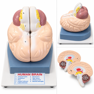
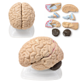
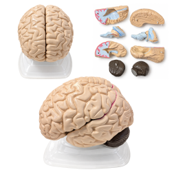
Human Brain Model, Life Size, 2 Parts, Hand-Painted with Numbered Structures
Two-part anatomical brain with numbered structures and hand-painted details.SKU: BRAIN1Human Brain Model, Life Size, 2 Parts, Hand-Painted with Numbered Structures
This anatomical teaching model illustrates the external and internal organization of the human brain through a vertically divided, two-part design. The detachable halves reveal selected internal structures and their spatial relationships, while the exterior reproduces the characteristic gyri and sulci of the cerebral cortex. Hand-painted anatomical details use contrasting colors to distinguish major brain regions. Numbered structures correspond to a reference key, allowing individual features to be identified during anatomical examination. The sectional construction provides direct access to internal anatomy while preserving the overall form of the assembled brain.
Product Specifications
SKU BRAIN1 Product Type Human Brain Anatomical Model Scale Life-Size (1:1) Number of Parts 2 Detachable Halves Section Type Vertical / Midsagittal Division Finish Hand-Painted Anatomical Details Anatomical Identification Numbered Structures with Reference Key Featured Structures Frontal Lobe, Cerebral Hemispheres, Cerebral Cortex, Cerebellum, Pons, Corpus Callosum, Lateral Ventricle, Optic Nerve, Medulla Oblongata, Spinal Cord Surface Anatomy Molded Cerebral Gyri and Sulci Display Base Tray-Style Display Stand
Applications and Benefits:
- Demonstrates external and internal human brain anatomy in biology and anatomy classrooms.
- Allows hands-on examination of two detachable anatomical halves.
- Supports identification of major cerebral structures through numbered references and an anatomical key.
- Illustrates the spatial relationships between the cerebral hemispheres, cerebellum, brainstem, and selected internal structures.
- Provides a visual reference for studying cerebral gyri, sulci, and sectional brain anatomy.
- Supports medical and nursing education through anatomical identification exercises.
- Useful for introductory neuroscience and psychology instruction involving the central nervous system.
- Suitable for homeschool science education and independent anatomy study.
- Provides an educational display for science museums, classrooms, and teaching laboratories.
- Includes a tray-style stand for supporting the assembled model during presentation and examination.
Caution:
For educational and demonstration purposes only. Not intended for medical diagnosis, clinical procedures, or treatment. Anatomical colors and numbered references are intended to facilitate identification and may simplify certain structures. Handle detachable sections carefully to avoid damage to connection points and painted surfaces. Clean with a soft, slightly damp cloth and avoid abrasive cleaners or strong solvents.
- Availability: In Stock
- Model: BRAIN1
-
Brand:

- 2 parts
- anatomical
- anatomy
- anatomy model
- biology
- brain
- BRAIN 1
- brain anatomy
- brain halves
- brain model
- brain models
- brain sections
- brain teaching
- BRAIN1
- brains
- brainstem
- cerebellum
- cerebral
- cerebrum
- corpus callosum
- demonstration
- dissectible
- education
- frontal lobe
- hand painted
- human brain
- laboratory
- lateral ventricle
- life size
- life-size
- medical
- medulla
- midsagittal
- nervous system
- neuroanatomy
- neurology
- neuroscience
- numbered anatomy
- numbered brain
- nursing
- optic nerve
- pons
- reference key
- sagittal
- sectional
- spinal cord
- teaching
- two parts
-