
-
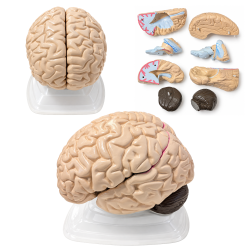 3307-2Human Brain Anatomical Model, 8 Parts, Vintage, with Internal Structures$69
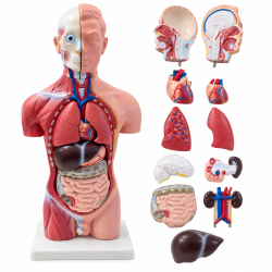 ACMT3Male Human Torso Anatomical Model, 16.5 Inches,$125
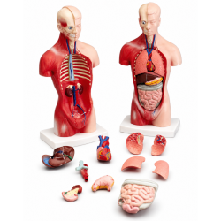 ACMT4Human Torso Anatomy Model, 10 1/8 in$12
 SKELETON_16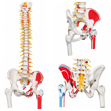
Flexible Human Spine Anatomical Model, Life Size.
Spinal column with movable femur heads and painted muscle attachments.SKU: 3394-1Deluxe Flexible Human Spine Anatomical Model, Life Size, with Femur Heads and Painted Muscles
This deluxe anatomical teaching model reproduces the human vertebral column with associated skeletal, nervous, vascular, and muscular attachment structures. Its flexible spinal mounting permits controlled movement to demonstrate natural spinal curvatures and the relationships between adjacent vertebrae. Articulated femoral heads provide an additional representation of the connection between the pelvis, hip joints, and spinal column. Muscle attachment sites are identified through contrasting painted markings, with red indicating muscle origins and blue indicating muscle insertions. The model also illustrates an intervertebral disc prolapse and selected neurological and vascular features, including spinal nerve exits, the cauda equina, cervical vertebral artery, and exposed brainstem. These anatomical details allow examination of spinal structure, vertebral alignment, and musculoskeletal relationships.
Product Specifications
SKU 3394-1 Product Type Flexible Human Spine Anatomical Model with Pelvis and Femur Heads Anatomical Scale Life-Size (1:1) Height Approximately 83 cm (32.7 inches) Weight Approximately 2.1 kg (4.63 lb) Spinal Construction Flexible Spinal Mounting Vertebral Regions Cervical, Thoracic, Lumbar, Sacrum, Coccyx Pelvis Male Pelvis Femur Heads Movable, Included Muscle Origin Markings Red-Painted Attachment Sites Muscle Insertion Markings Blue-Painted Attachment Sites Disc Condition Illustrated L3-L4 Intervertebral Disc Prolapse Nervous System Details Spinal Nerve Exits, Cauda Equina, Exposed Brainstem Representation Vascular Anatomy Cervical Vertebral Artery Additional Structures Intervertebral Discs, Occipital Plate, Sacral Opening Display Stand Not Included
Applications and Benefits:
- Demonstrates the complete human vertebral column and its major anatomical regions.
- Allows hands-on demonstrations of spinal curvature, flexibility, positioning, and vertebral alignment.
- Supports identification of cervical, thoracic, and lumbar vertebrae, as well as the sacrum and coccyx.
- Illustrates muscle attachment sites through red-painted origins and blue-painted insertions.
- Demonstrates the anatomical relationship between the vertebral column, male pelvis, and movable femoral heads.
- Provides a visual example of an L3-L4 intervertebral disc prolapse for introductory spinal pathology education.
- Supports examination of spinal nerve exits, the cauda equina, and associated neurological structures.
- Illustrates the anatomical location of the cervical vertebral artery and exposed brainstem representation.
- Supports the study of musculoskeletal anatomy, spinal biomechanics, and joint relationships.
- Suitable for medical schools, nursing programs, biology classrooms, and anatomy laboratories.
- Provides a teaching reference for physical therapy, physiotherapy, chiropractic, and orthopedic education.
- Useful for healthcare demonstrations and independent anatomical study.
Caution:
For educational and demonstration purposes only. Not intended for medical diagnosis, clinical procedures, treatment, or patient-specific biomechanical assessment. Spinal movements and anatomical features are illustrative and may not reproduce the full range or mechanical behavior of the human vertebral column. The L3-L4 disc prolapse is a simulated anatomical condition. Flex the model gently and avoid excessive bending, twisting, or pulling on the vertebrae, movable femur heads, spinal nerves, and other projecting structures. A display stand is not included.
- Availability: In Stock
- Model: 3394-1
-
Brand:

- 3394 1
- 3394-1
- 33941
- anatomical
- anatomy
- anatomy model
- backbone
- biology
- biomechanics
- brainstem
- cauda equina
- cervical spine
- chiropractic
- coccyx
- deluxe spine
- demonstration
- disc prolapse
- education
- femur
- femur heads
- flexible spine
- herniated disc
- hip joints
- human spine
- intervertebral discs
- L3-L4
- laboratory
- life size
- lumbar spine
- male pelvis
- medical
- muscle attachments
- muscle insertions
- muscle origins
- musculoskeletal
- nerve roots
- nursing
- occipital plate
- orthopedics
- pelvic anatomy
- physical therapy
- physiotherapy
- sacrum
- spinal anatomy
- spinal column
- spinal nerves
- spine
- spine anatomy
- spine model
- spine models
- spines
- teaching
- thoracic spine
- vertebra
- vertebrae
- vertebral artery
- vertebral column
-
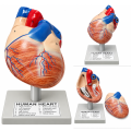 Human Heart Anatomical Model, 2 Parts, Hand-Painted with Numbered Structures
-
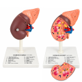 Human Kidney Anatomical Model with 16 Labeled Structures
-
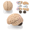 Human Brain Anatomical Model, 8 Parts, Vintage, with Internal Structures
-
 Skin Anatomy Model
-
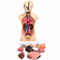 Human Torso Model, Male/Female 18" (45 Cm)
-
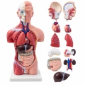 Male Human Torso Anatomical Model, 16.5 Inches,
-
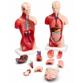 Human Torso Anatomy Model, 10 1/8 in
-
 Atomic Model Set Laboplast
-
 Atomic Model Set Junior Code 58002 Mark
-
 Human Eye Anatomical Model, 6X Enlarged, 6 Parts, Hand-Painted with Numbered Structures
-
 Model Of The Ear


