
-
 ATOMSETLPAtomic Model Set Laboplast$60
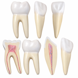 TEETH305Human Teeth Anatomy Model Set - 12× Enlarged, 3 Tooth Types$65
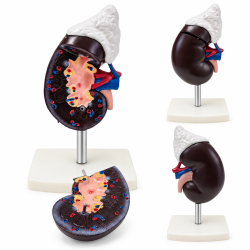 KIDNEY310-3Human Kidney and Adrenal Gland Model - Life-Size, 2 Parts$35
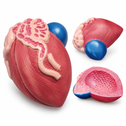 BLADDER317Male Bladder and Prostate Anatomy Model - Median Section$45
 SKELETON16Human Skeleton Model 16-Inch$25 - $432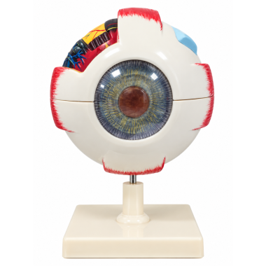
Human Eye Anatomical Model, 6X Enlarged, 6 Parts, Hand-Painted with Numbered Structures
Six detachable PVC sections with 12 numbered anatomical features.SKU: E3304Human Eye Anatomical Model, 6X Enlarged, 6 Parts, Hand-Painted with Numbered Structures
This enlarged anatomical teaching model illustrates the external and internal organization of the human eye through a detailed sectional design. Its detachable components reveal the eye's protective layers, optical structures, and internal anatomy, allowing individual features to be examined and their spatial relationships understood. Hand-painted anatomical details use contrasting colors to distinguish the sclera, choroid, retina, iris, lens, vitreous body, and associated structures. Numbered identification references correspond to twelve major anatomical features, including the optic nerve, central fovea, ciliary body, and ocular blood vessels. The dissectible construction allows the components to be removed and reassembled, providing a direct visual representation of the eye's anatomical organization.
Product Specifications
SKU E3304 Product Type Human Eye Anatomical Model Anatomical Scale 6X Life-Size (6:1) Number of Parts 6 Detachable Components Primary Material PVC (Polyvinyl Chloride) Surface Finish Hand-Painted Anatomical Details Anatomical References 12 Numbered Features Featured Structures Cornea, Sclera, Choroid, Retina, Iris, Lens, Vitreous Body, Optic Nerve, Central Fovea, Ciliary Body, Ocular Blood Vessels Dimensions Approximately 8.07 × 7.87 × 5.71 inches Weight Approximately 1.32 lb Construction Type Sectional, Dissectible Display Base Removable Display Stand
Anatomical Structures Identified
- 1. Cornea
- 2. Sclera
- 3. Choroid
- 4. Retina
- 5. Iris
- 6. Lens
- 7. Vitreous Body
- 8. Optic Nerve
- 9. Central Fovea
- 10. Choroidal Blood Vessels
- 11. Ciliary Body
- 12. Central Retinal Artery and Vein
Applications and Benefits:
- Demonstrates the external and internal anatomy of the human eye in biology and anatomy classrooms.
- Provides a six-times-enlarged representation for examining detailed ocular structures.
- Allows hands-on examination of six detachable anatomical components.
- Supports identification of twelve major anatomical features through numbered references.
- Illustrates the organization of the sclera, choroid, and retina as the principal layers of the eyeball.
- Demonstrates the relationship between the cornea, iris, lens, and other structures involved in vision.
- Supports examination of the optic nerve, central fovea, ciliary body, and ocular blood vessels.
- Suitable for introductory ophthalmology, optometry, and vision science instruction.
- Supports medical, nursing, and healthcare education through anatomical identification exercises.
- Provides a physical reference for independent study and educational laboratory demonstrations.
- Suitable for science museums, classrooms, and educational displays.
- Includes a removable display stand for supporting the assembled model during presentations and storage.
Caution:
For educational and demonstration purposes only. Not intended for medical diagnosis, clinical procedures, or treatment. Anatomical structures, colors, and proportions may be simplified for instructional clarity. Handle detachable components carefully to prevent damage to connection points and painted surfaces. Clean with a soft, slightly damp cloth and avoid abrasive cleaners or strong solvents.
- Availability: In Stock
- Model: E3304
-
Brand:

- 12 structures
- 6 parts
- 6X enlarged
- 6X eye
- anatomical
- anatomy
- anatomy model
- biology
- blood vessels
- choroid
- ciliary body
- cornea
- demonstration
- detachable
- dissectible
- E 3304
- E3304
- education
- enlarged eye
- eye
- eye anatomy
- eye dissection
- eye layers
- eye model
- eye models
- eyeball
- eyes
- fovea
- hand painted
- human eye
- iris
- laboratory
- lens
- medical
- numbered anatomy
- numbered eye
- nursing
- ocular
- ocular anatomy
- ophthalmology
- optic nerve
- optometry
- PVC
- retina
- retinal artery
- retinal vein
- sclera
- six parts
- teaching
- vision
- visual system
- vitreous
-
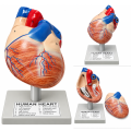 Human Heart Anatomical Model, 2 Parts, Hand-Painted with Numbered Structures
-
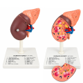 Human Kidney Anatomical Model with 16 Labeled Structures
-
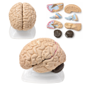 Human Brain Anatomical Model, 8 Parts, Vintage, with Internal Structures
-
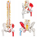 Flexible Human Spine Anatomical Model, Life Size.
-
 Skin Anatomy Model
-
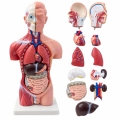 Male Human Torso Anatomical Model, 16.5 Inches,
-
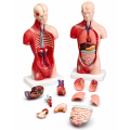 Human Torso Anatomy Model, 10 1/8 in
-
 Atomic Model Set Laboplast
-
 Atomic Model Set Junior Code 58002 Mark
-
 Model Of The Ear
-
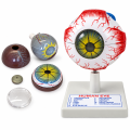 Human Eye Anatomical Model, 3X Enlarged, 6 Parts, with Numbered Structures


