
-
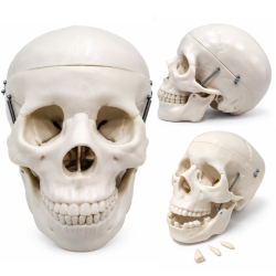 SKULL104Human Skull Model - Life-Size, 3 Parts, Removable Teeth$39 - $320
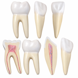 TEETH305Human Teeth Anatomy Model Set - 12× Enlarged, 3 Tooth Types$65
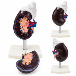 KIDNEY310-3Human Kidney and Adrenal Gland Model - Life-Size, 2 Parts$35
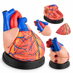 HEART307C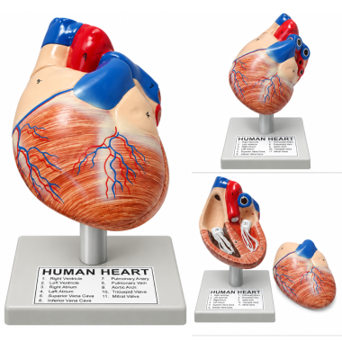
Human Heart Anatomical Model, 2 Parts, Hand-Painted with Numbered Structures
Dissectible heart with internal structures and numbered identification key.SKU: 1020-33Human Heart Anatomical Model, 2 Parts, Hand-Painted with Numbered Structures
This dissectible human heart model illustrates the relationship between the heart's external vascular anatomy and internal cardiac structures. A removable heart wall section exposes the ventricular chambers, valves, and chordae tendineae for direct examination. The hand-painted exterior distinguishes the cardiac muscle, coronary blood vessels, and major vascular connections using contrasting anatomical colors. Numbered reference points correspond to an identification key printed directly on the display base, allowing anatomical structures to be identified without a separate reference chart. The model is mounted on a rigid support that holds the heart in position during observation and disassembly.
Product Specifications
SKU 1020-33 Anatomical Subject Human Heart Model Configuration Dissectible Number of Parts 2 Primary Material Molded Polymer Surface Finish Hand-Painted Removable Component Heart Wall Section Numbered Anatomical Structures 11 Identification System Numbered Structures with Integrated Reference Key Display Base Gray Plastic Stand with Vertical Support
Applications and Benefits:
- Demonstrates external and internal anatomy of the human heart in biology and anatomy classrooms.
- Supports medical, nursing, and healthcare education through three-dimensional examination of cardiac structures.
- Illustrates the relationship between the right and left atria, ventricles, and major blood vessels.
- Allows observation of the tricuspid and mitral valves and associated chordae tendineae.
- Identifies the superior vena cava, inferior vena cava, pulmonary artery, pulmonary vein, and aortic arch.
- Demonstrates coronary blood vessel distribution and the external muscular structure of the heart.
- Provides 11 numbered anatomical references: right ventricle, left ventricle, right atrium, left atrium, superior vena cava, inferior vena cava, pulmonary artery, pulmonary vein, aortic arch, tricuspid valve, and mitral valve.
- Supports identification exercises using the reference key printed on the display base.
- Allows repeated removal and reassembly of the heart wall during laboratory demonstrations.
- Supports independent anatomical study and introductory cardiology instruction.
Caution:
Intended for educational and demonstration purposes only. Not designed for medical diagnosis or clinical procedures. Handle removable components carefully to avoid damage to anatomical details and mounting connections. Do not force parts during disassembly or reassembly.
- Availability: In Stock
- Model: 1020-33
-
Brand:

- 1020 33
- 1020-33
- 102033
- anatomical
- anatomy
- anatomy model
- aorta
- aortic arch
- arteries
- atria
- atrium
- biology
- blood vessels
- cardiac
- cardiac model
- cardiology
- cardiovascular
- chordae tendineae
- circulation
- classroom
- coronary
- dissectible
- dissection
- educational
- handpainted
- heart
- heart anatomy
- heart chambers
- heart model
- heart valves
- heart1020
- hearts
- human heart
- laboratory
- medical
- medical model
- mitral valve
- myocardium
- nursing
- pericardium
- polymer
- pulmonary
- removable
- simulation
- teaching
- teaching model
- tricuspid valve
- valves
- veins
- vena cava
- ventricle
- ventricles
-
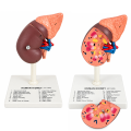 Human Kidney Anatomical Model with 16 Labeled Structures
-
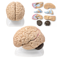 Human Brain Anatomical Model, 8 Parts, Vintage, with Internal Structures
-
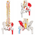 Flexible Human Spine Anatomical Model, Life Size.
-
 Skin Anatomy Model
-
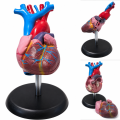 Human Heart Anatomical Model, Life Size, 2 Parts, Budget Edition
-
 Human Torso Model, Male/Female 18" (45 Cm)
-
 Male Human Torso Anatomical Model, 16.5 Inches,
-
 Human Torso Anatomy Model, 10 1/8 in
-
 Atomic Model Set Laboplast
-
 Atomic Model Set Junior Code 58002 Mark
-
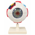 Human Eye Anatomical Model, 6X Enlarged, 6 Parts, Hand-Painted with Numbered Structures


