
-
 4DSKINSkin Anatomy Model$32
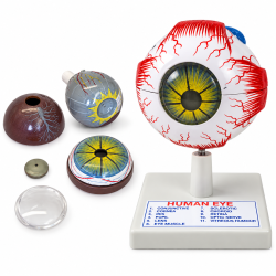 MODELEYEHuman Eye Anatomical Model, 3X Enlarged, 6 Parts, with Numbered Structures$39
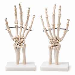 HAND114Human Hand Skeleton Model - Life-Size with Stand$24
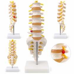 SPINE119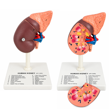
Human Kidney Anatomical Model with 16 Labeled Structures
Kidney model with 16 identified renal and vascular structures.SKU: 1020-55Human Kidney Anatomical Model with 16 Labeled Structures
This anatomical teaching model illustrates the external and internal organization of the human kidney, including its associated blood vessels, adrenal gland, and urinary drainage structures. The anatomical representation exposes the renal cortex, medullary pyramids, renal columns, papillae, calyces, and renal pelvis, showing how these structures are arranged within the kidney. The renal artery and vein are represented alongside the suprarenal vessels, demonstrating their relationship to the kidney and adrenal gland. Sixteen identified anatomical features provide a reference for distinguishing major renal structures and understanding the connections between the kidney's internal anatomy, vascular supply, and urine collection system.
Product Specifications
SKU 1020-55 Product Type Human Kidney Anatomical Model Anatomical References 16 Identified Structures Featured Organs Kidney and Adrenal (Suprarenal) Gland Renal Parenchyma Renal Cortex, Medullary Pyramids, Renal Papillae, Renal Columns Urinary Collection Structures Minor Calyces, Major Calyces, Renal Pelvis, Ureter Vascular Anatomy Renal Artery, Renal Vein, Right Suprarenal Artery, Right Suprarenal Vein External Landmarks Upper Pole, Lower Pole, Renal Hilum
Anatomical Structures Identified
- 1. Suprarenal (Adrenal) Gland
- 2. Right Suprarenal Vein
- 3. Right Suprarenal Artery
- 4. Renal Artery
- 5. Renal Vein
- 6. Renal Pelvis
- 7. Ureter
- 8. Upper Pole of Kidney
- 9. Lower Pole of Kidney
- 10. Renal Hilum
- 11. Renal Cortex
- 12. Medullary Pyramid
- 13. Renal Papilla
- 14. Renal Column
- 15. Minor Calyx
- 16. Major Calyx
Applications and Benefits:
- Demonstrates the external and internal anatomy of the human kidney in biology and anatomy classrooms.
- Supports identification of 16 renal, vascular, and associated anatomical structures.
- Illustrates the organization of the renal cortex, medullary pyramids, papillae, and renal columns.
- Demonstrates the pathway of urine collection from the renal papillae through the calyces, renal pelvis, and ureter.
- Supports the study of renal circulation through the identification of major renal blood vessels.
- Illustrates the anatomical relationship between the kidney and suprarenal gland.
- Provides a visual reference for studying the renal hilum and associated anatomical structures.
- Suitable for introductory nephrology and human urinary system education.
- Supports medical and nursing education through anatomical identification exercises.
- Provides a physical reference for independent anatomical study and educational laboratory demonstrations.
- Suitable for science museums, educational displays, and classroom presentations.
Caution:
For educational and demonstration purposes only. Not intended for medical diagnosis, clinical procedures, or treatment. Anatomical structures may be simplified or enlarged for instructional clarity and may not reproduce every detail of human renal anatomy. Handle the model carefully to avoid damage to projecting blood vessels and anatomical structures. Clean with a soft, slightly damp cloth and avoid abrasive cleaners or strong solvents.
- Availability: In Stock
- Model: 1020-55
-
Brand:

- 1020 55
- 1020-55
- 102055
- 16 structures
- adrenal gland
- anatomical
- anatomy
- anatomy model
- biology
- demonstration
- education
- human kidney
- kidney
- kidney anatomy
- kidney model
- kidney models
- kidney structures
- kidneys
- labeled kidney
- laboratory
- major calyx
- medical
- minor calyx
- nephrology
- nursing
- renal
- renal anatomy
- renal artery
- renal circulation
- renal column
- renal cortex
- renal hilum
- renal medulla
- renal papilla
- renal pelvis
- renal pyramids
- renal vein
- science model
- suprarenal gland
- teaching
- ureter
- urinary system
- urinary tract
-
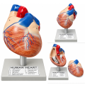 Human Heart Anatomical Model, 2 Parts, Hand-Painted with Numbered Structures
-
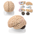 Human Brain Anatomical Model, 8 Parts, Vintage, with Internal Structures
-
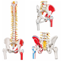 Flexible Human Spine Anatomical Model, Life Size.
-
 Skin Anatomy Model
-
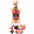 Human Torso Model, Male/Female 18" (45 Cm)
-
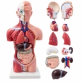 Male Human Torso Anatomical Model, 16.5 Inches,
-
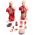 Human Torso Anatomy Model, 10 1/8 in
-
 Atomic Model Set Laboplast
-
 Atomic Model Set Junior Code 58002 Mark
-
 Human Eye Anatomical Model, 6X Enlarged, 6 Parts, Hand-Painted with Numbered Structures
-
 Model Of The Ear


